Scientists may have found why diabetic wounds refuse to heal
Chronic diabetic wounds may fail to heal because immune cells remain locked in damaging inflammatory states instead of transitioning toward tissue repair. Researchers are exploring treatments that reprogram these immune responses, raising hopes for more targeted and effective therapies for diabetic ulcers.
Key points
- More than 131 million people worldwide have chronic diabetic ulcers, costing about $755 billion yearly.
- A review examines how immune cells like macrophages and neutrophils behave in diabetic wounds.
- Therapies targeting immune cells at specific healing stages may improve treatment.
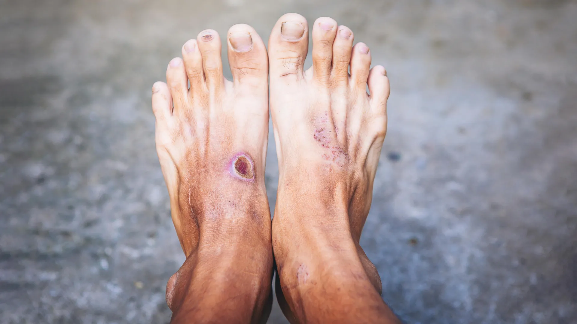
Chronic diabetic ulcers affect more than 131 million people worldwide and are linked to roughly $755 billion in healthcare costs each year. They carry serious risks, including amputation and death, and are often resistant to healing.
A comprehensive review by Yi Ru and colleagues examines how different immune cell populations change over time during diabetic wound healing. The review covers monocytes, macrophages, dendritic cells, neutrophils, mast cells, B cells, T cells, and natural killer cells, focusing on where these cells appear and how diabetes disrupts their normal behavior.
Immune cell dysfunction in diabetic wounds
Monocytes and macrophages receive particular attention. In normal healing, monocytes enter damaged tissue and become macrophages that shift from pro-inflammatory M1 states to pro-reparative M2 states. In diabetic wounds, this transition can break down, leaving macrophages locked in pro-inflammatory states that prolong damage.
Neutrophils, among the first responders to injury, can have poorly regulated neutrophil extracellular trap formation in diabetic wounds, prolonging inflammation. Mast cell degranulation differs substantially between normal and diabetic wounds, and excessive activation may sustain chronic inflammation. Dendritic cells become less effective at efferocytosis, allowing dead cellular material to accumulate and trigger inflammation, with reduced SLC7A11 transporter activity implicated.
a better understanding of how immune cell populations interact over time could lead to more precise and effective treatments for diabetic ulcers
— the authors
T cells, B cells and NK cells
Regulatory T cells, which normally suppress excessive immune activity and support tissue remodeling, decline in number and function in diabetic wounds, slowing healing. Dendritic epidermal T cells, a type of gamma-delta T cell in the skin, show lower activation and reduced cytokine production under diabetic conditions.
B cells and natural killer cells are less studied but emerging evidence suggests they contribute to healing. B cells can influence macrophage polarization, and recent evidence shows their recruitment can encourage M2 states to reduce inflammation.
Potential treatments and timing
New treatments could reprogram immune responses. The review highlights topical anti-cytokine biologics, drugs that promote macrophage polarization, mesenchymal stem cell therapies, extracellular vesicles, advanced biomaterials, smart dressings, Janus liposozyme technologies, macrophage-regulating drugs, and IL-15 superagonists.
The researchers emphasize that effective treatment may depend on when immune cells are targeted. Diabetic wounds pass through inflammatory, proliferative, and remodeling stages, each with a different immune environment. Future approaches could include personalized treatment plans based on immune profiling and combination therapies targeting multiple cell populations.