Stanford’s new microscope reveals living cells like never before
Stanford scientists have developed a revolutionary microscope that reveals the hidden inner workings of living cells in real time, without the fluorescent labels that can interfere with observations. The breakthrough could provide new insights into cancer, infectious diseases, and how drugs interact with cells.
Key points
- Stanford researchers built a label-free microscope, iISM, that images living cells at about 120 nanometers resolution.
- The system combines interferometric scattering microscopy with detector arrays from advanced confocal microscopes.
- Planned projects include cancer drug entry, plant-microbe interactions and malaria effects on red blood cells.
Researchers at Stanford University have developed a microscope that lets scientists watch structures inside living cells move and interact in real time without the fluorescent labels usually needed to make them visible. The instrument, called Interferometric Image Scanning Microscopy (iISM), reaches a resolution of approximately 120 nanometers, which the team describes as an unprecedented level of detail for a label-free approach. The work was described in the Nature portfolio journal Light: Science and Applications.
The microscope combines two advanced imaging techniques. According to the article, it could support work in cancer research, drug development, infectious disease studies and research on how plants interact with microorganisms.
Why labels are avoided
Many existing methods use fluorescent substances to highlight cell structures, but researchers can generally label only a small number of structures at once, making it hard to see how components across a whole cell interact. Fluorescent signals can also fade during observation, a process called photobleaching, and some markers may alter the behavior of the structures being studied.
The Stanford system images cells without such labels, allowing many components to be followed over extended periods in a more natural setting. It also needs substantially less illumination power than comparable label-free methods, reducing the risk of photodamage to living cells. The researchers say the technology is meant to complement fluorescence microscopy, not replace it.
It's a wonderful look into these complex little cellular boxes that drive our life.
— W.E. Moerner
How the instrument works
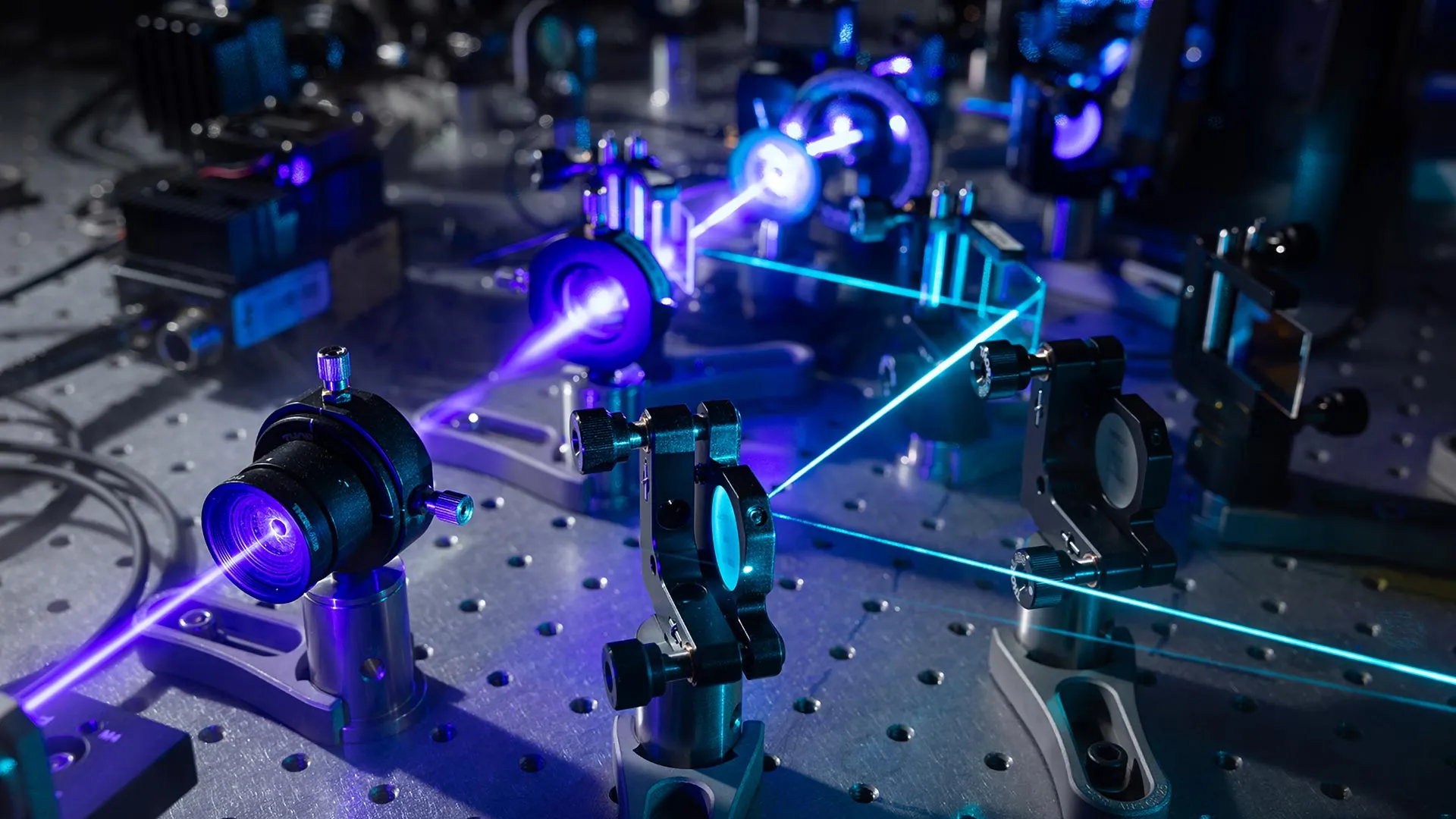
The design merges interferometric scattering microscopy, which detects light scattered by tiny structures, with technology adapted from advanced confocal microscopes. A laser illuminates the cell, and a second laser beam amplifies the weak scattered signal through interference. An array detector collects tens to hundreds of views of the same region, and a computational method combines them into sharper images with depth information.
Senior author W.E. Moerner, the Harry S. Mosher Professor of Chemistry, said the instrument offers a new way to see cellular activity. "It's a wonderful look into these complex little cellular boxes that drive our life," he said.
Early research collaborations
Three collaborations with other Stanford groups are underway or in development. One observes interactions among plant cells, fungi and bacteria in real time. Another examines how a cancer drug enters a cell. A third, still being planned, will study how red blood cells change shape when exposed to malaria infection. First author Michelle Kueppers said the technique has broad applications beyond a single field.