A revolutionary new microscope could reveal the hidden machinery of life
Researchers have developed a revolutionary microscope that uses an incredibly powerful laser to make previously invisible cellular structures easier to see. The technology dramatically improves image clarity, allowing scientists to examine small proteins that conventional electron microscopes struggle to capture. It could eventually reveal how tiny molecular machines interact inside cells, providing an unprecedented…
Key points
- Physicists at UC Berkeley have adapted phase-contrast microscopy to electron microscopy using a device called a laser phase plate.
- The laser phase plate improved resolution for six test samples and allowed imaging of proteins as small as 50 kilodaltons, below the conventional cryo-EM limit of about 70 kilodaltons.
- Müller named the upgraded microscope Theia, and Biohub is building a related two-laser instrument in Redwood City, California.
Physicists at the University of California, Berkeley, have developed a laser phase plate that brings phase-contrast imaging, a technique honored with a Nobel Prize in 1953, to electron microscopy. The device is designed to improve cryoelectron microscopy (cryo-EM) and cryoelectron tomography (cryo-ET), imaging methods used to study protein structures.
Cryo-EM has a major limitation: most human proteins are too small to be imaged clearly. The laser phase plate is intended to overcome this by increasing image contrast, potentially allowing scientists to image proteins as small as one-third the size of those that challenge current microscopes.
How the device works
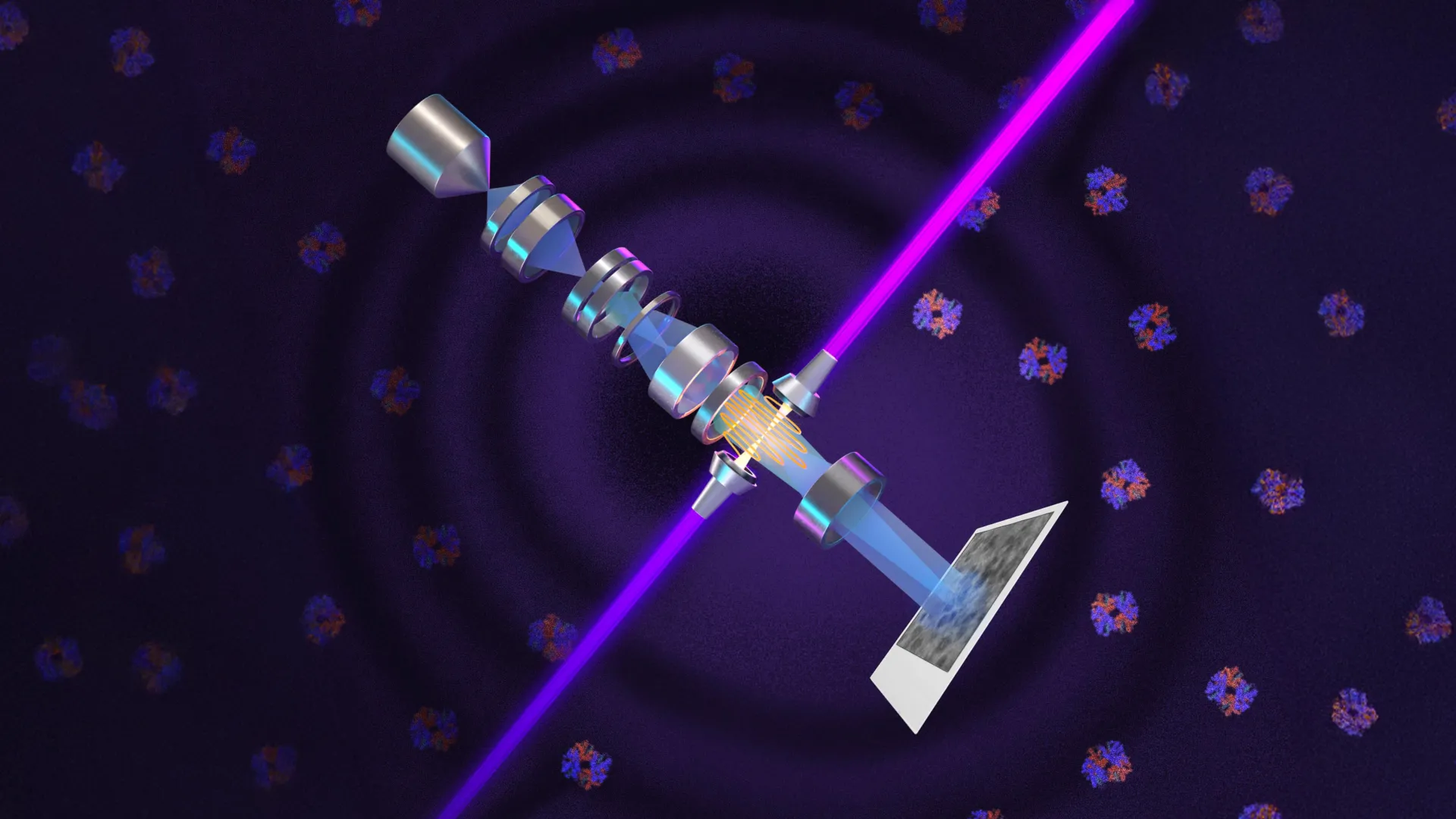
The system relies on what the researchers describe as the world's most intense focused continuous-wave laser. Instead of only illuminating a specimen, the laser interacts with the electron beam and changes its phase to boost contrast, making small molecules such as hemoglobin easier to distinguish from their surroundings.
Müller spent 15 years developing the concept after proposing it with Robert Glaeser in 2010. The laser is confined in a mirrored spherical chamber where it reflects more than 10,000 times, building intensity. Müller said it is "75 kilowatts focused to a few microns."
This technology is a step function change for biology
— Stephani Otte
Tests and next steps
The team tested the upgraded microscope, named Theia, on six samples of different sizes and preparation methods. The laser consistently improved resolution, with the largest gains for small or difficult specimens. The researchers reconstructed structures of aldolase and hemoglobin, with the improvement especially pronounced for the smaller hemoglobin.
Conventional cryo-EM struggles with proteins below about 70 kilodaltons, which account for roughly 90% of the human proteome. With the laser phase plate, proteins as small as 50 kilodaltons can now be imaged, though it remains challenging. Biohub, which provided funding, is building a related microscope with two lasers at its Redwood City, California, facility. Both teams are working with Thermo Fisher Scientific.